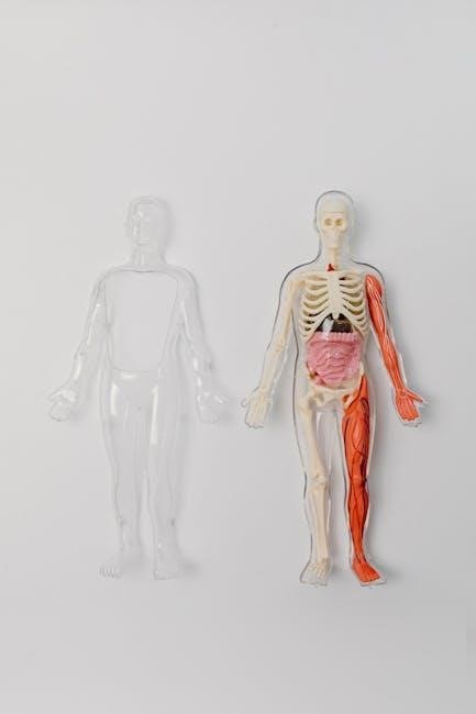
Welcome to the Human Anatomy and Physiology Lab Manual, a comprehensive guide designed to enhance your understanding of the human body through hands-on experimentation and detailed exploration. This manual provides a structured approach to learning, combining theoretical knowledge with practical lab experiences to ensure a deep grasp of essential concepts. By engaging in exercises and activities, you will gain valuable insights into the structure and function of key body systems, preparing you for further study and real-world applications in healthcare and science. This resource is tailored to support your academic journey, fostering critical thinking and scientific inquiry.
1.1 Importance of Lab Work
Lab work is essential for gaining hands-on experience in human anatomy and physiology. It allows students to apply theoretical knowledge in practical scenarios, enhancing understanding of complex biological processes. Through dissections, observations, and experiments, learners develop critical thinking and problem-solving skills. Labs also foster scientific inquiry and collaboration, preparing students for real-world applications in healthcare and research.
- Enhances comprehension of anatomical structures and physiological functions;
- Develops practical skills necessary for future careers.
- Encourages curiosity and deeper exploration of biological systems.
Lab experiences create a foundation for lifelong learning in the sciences.
1.2 Lab Objectives and Outcomes
The primary objective of lab work is to provide a comprehensive understanding of human anatomy and physiology through hands-on activities. Students will identify and examine anatomical structures, conduct physiological experiments, and analyze data. Key outcomes include improved observation skills, enhanced critical thinking, and the ability to interpret scientific results. These experiences prepare students for professional applications in healthcare and research.
- Develop proficiency in using lab equipment and tools.
- Understand the interrelationships between body systems.
- Cultivate skills in scientific inquiry and experimentation;
Lab outcomes emphasize practical application and real-world relevance.
Preparing for the Lab
Preparing for the lab involves reviewing assigned materials, organizing necessary supplies, and familiarizing oneself with lab protocols to ensure efficiency and safety during experiments.
- Review notes and lab manuals beforehand.
- Gather required tools and personal protective equipment.
- Adhere to dress code and safety guidelines.
2.1 Safety Protocols
Safety protocols are crucial to prevent accidents and ensure a secure environment. Proper PPE, including gloves and goggles, must be worn. Handling biological materials and equipment requires caution, and emergency procedures like fire extinguisher locations and first aid kits must be known. Adhering to specific guidelines helps prevent injuries and maintains a safe workspace for all participants.
2.2 Essential Equipment and Tools
Essential equipment includes microscopes, dissecting tools, and measurement devices. Gloves, lab coats, and goggles are also vital. Specimen trays, scalpels, and forceps are used for dissections. Thermometers, pH indicators, and scales aid in physiological measurements. Proper storage and organization of tools ensure efficiency and safety during lab activities, supporting accurate observations and successful experiments.
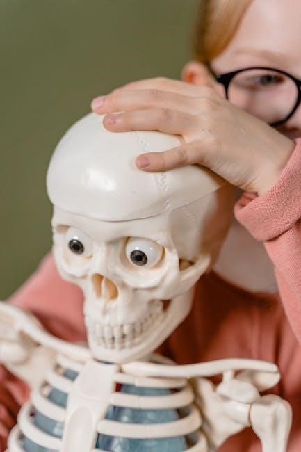
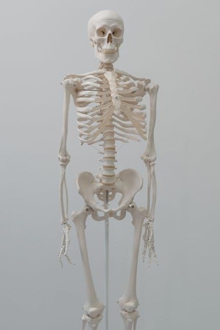
Skeletal System
The skeletal system provides structural support and protection, comprising 206 bones in the adult human body. It includes long bones, short bones, and flat bones, facilitating movement and housing vital organs.
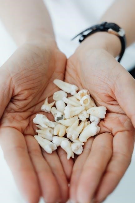
3.1 Bone Structure Analysis
Bone structure analysis involves examining the composition and organization of bones. The diaphysis (shaft) and epiphysis (end) are key regions. The metaphysis connects these, containing spongy bone. Compact bone forms the outer layer, while spongy bone is porous. The medullary cavity houses bone marrow. This structure provides strength, supports movement, and protects internal organs, illustrating the skeletal system’s functional design.
3.2 Joint Dissection
Joint dissection involves the examination of articulations between bones. Types include synovial, cartilaginous, and fibrous joints. The procedure requires careful dissection to expose the joint cavity, ligaments, and cartilage. Students identify and analyze the structural components, understanding their roles in movement and stability. This hands-on activity enhances comprehension of joint functionality and prepares for studying musculoskeletal disorders.
Muscular System
The muscular system comprises skeletal, smooth, and cardiac muscles. It facilitates movement, maintains posture, and regulates body functions; Studying this system enhances understanding of muscle structure, function, and disorders.
4.1 Muscle Tissue Identification
Muscle tissue identification involves analyzing skeletal, smooth, and cardiac muscles. Skeletal muscles are striated and attached to bones, enabling voluntary movement. Smooth muscles are non-striated and found in internal organs, functioning involuntarily. Cardiac muscles are striated with intercalated discs, ensuring synchronized heart contractions. Lab exercises include microscopic examination and histological staining to distinguish these tissues based on their unique structural and functional characteristics.
4.2 Physiological Responses
Physiological responses in muscles involve contraction and relaxation mechanisms. Skeletal muscles respond to nerve stimulation, exhibiting twitch, tetanus, and muscle fatigue. Smooth muscles react to hormones and stretching, maintaining muscle tone. Cardiac muscles have intrinsic rhythmic contractions regulated by electrolytes like calcium and potassium. Lab experiments measure these responses using techniques like electromyography to record muscle activity and understand neuromuscular junctions, providing vital insights into muscle physiology and its clinical relevance in diagnosing and treating muscle-related disorders.
Nervous System
The nervous system is a complex control and communication network. It integrates and processes sensory information, enabling motor responses. Key components include the brain, spinal cord, and peripheral nerves, which transmit signals through neurons and synapses. Lab activities focus on exploring nerve structure, reflexes, and neural function, enhancing understanding of nervous system physiology and its role in human health.
5.1 Neuron Structure
A neuron, or nerve cell, is the functional unit of the nervous system. It consists of dendrites, a cell body, and an axon. Dendrites receive signals, while the axon transmits them to other neurons or tissues. The cell body contains the nucleus and organelles essential for protein synthesis. Lab activities involve studying neuron morphology under microscopes and exploring how structure relates to function in neural communication and signaling.
5.2 Reflex Arc Demonstrations
Reflex arcs are demonstrated to illustrate the neural pathway for involuntary responses. Labs involve simulating reflex actions, such as knee-jerk reactions, to observe the sequence of events. Students analyze the roles of sensory neurons, the spinal cord, motor neurons, and effectors. Activities emphasize understanding neural integration and the importance of reflexes in maintaining homeostasis and responding to stimuli.
Circulatory System
The circulatory system, comprising the heart, blood, and blood vessels, is vital for transporting oxygen, nutrients, and hormones throughout the body, maintaining homeostasis and overall health.
6.1 Blood Typing
Blood typing is a fundamental lab procedure used to classify blood based on the presence or absence of specific antigens on red blood cells. The ABO blood group system and Rh factor are commonly tested. Students learn to use antiserum to determine reactions, identifying types A, B, AB, or O, and Rh positive or negative. This exercise emphasizes compatibility for transfusions and pregnancy. Proper safety protocols are essential when handling blood samples.
- Universal donor: O-
- Universal recipient: AB+
6.2 Heart Dissection
Heart dissection involves carefully examining the structure and function of the heart. Students learn to identify chambers, valves, and blood vessels. The process begins with an exterior examination, followed by precise incisions to expose internal structures. Key observations include the thickness of ventricular walls and the condition of valves. This hands-on activity enhances understanding of blood flow and cardiac function.
Respiratory System
The respiratory system section focuses on understanding breathing mechanisms and gas exchange. Labs include lung capacity measurements and airway structure examinations to explore respiratory functions and their physiological significance.
7.1 Lung Capacity
Lung capacity measures the total amount of air the lungs can hold, essential for assessing respiratory health. Students learn to measure vital capacity using spirometry, recording maximum exhalation after full inhalation. Proper breathing techniques and calibration ensure accurate results. This exercise highlights individual variations due to age, sex, height, and health conditions, emphasizing the importance of respiratory function in overall physiology.
7.2 Trachea and Bronchi
The trachea, or windpipe, is a cartilage-reinforced tube connecting the larynx to the bronchi. It divides into primary bronchi, each leading to a lung. Histological examination reveals ciliated epithelium and mucous membranes. In lab, students examine tracheal and bronchial tissues, identifying structures like cartilage rings and glands. Dissection highlights airway branching and mucus clearance mechanisms, essential for respiratory function.
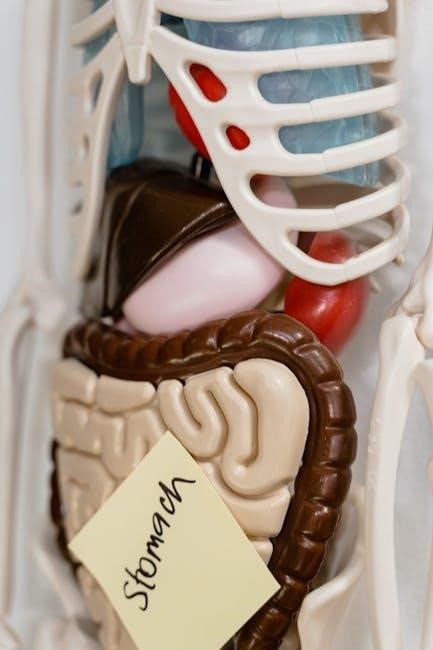
Digestive System
The digestive system includes the mouth, esophagus, stomach, small intestine, and large intestine. Each organ performs distinct functions like chewing, mixing with saliva, gastric digestion, nutrient absorption, and waste elimination. Enzymes play a crucial role in breaking down food into absorbable nutrients. Understanding the anatomy and physiology of digestion is essential for clinical applications and maintaining overall health.
8.1 Enzyme Functions
Enzymes are biological catalysts that accelerate chemical reactions in the body. They lower activation energy, enabling reactions to occur efficiently. In digestion, enzymes like amylase break down starch into sugars, while lipase targets fats. Lab exercises involve testing enzyme specificity and optimal conditions. Observing colorimetric assays helps determine enzyme activity levels. Understanding enzyme functions is crucial for grasping metabolic processes and diagnosing related disorders;
8.2 Organ Dissection
Organ dissection provides hands-on experience to explore internal structures and their functions. Students learn to identify key features of organs like the stomach, liver, and pancreas. Proper techniques are emphasized, including tool usage and workspace maintenance. Safety protocols, such as glove and goggles use, are essential. This exercise enhances understanding of anatomical relationships and prepares students for clinical applications.
Urinary System
The urinary system, involving kidneys, ureters, bladder, and urethra, is crucial for waste removal and fluid balance; Urinalysis and kidney structure studies provide essential insights into its functions.
9.1 Urinalysis
Urinalysis is a fundamental diagnostic tool in clinical settings, providing insights into urinary system health. It involves physical, chemical, and microscopic examinations of urine. Key parameters include color, clarity, pH, protein, glucose, ketones, blood, bilirubin, and urobilinogen levels. Abnormal findings, such as proteinuria or hematuria, can indicate kidney dysfunction, diabetes, or infections. Accurate testing aids in early disease detection and monitoring.
9.2 Kidney Structure
The kidney is a vital organ with a complex structure designed for filtration and waste removal. It consists of the renal cortex, renal medulla, and renal pelvis. Nephrons, the functional units, include the glomerulus and Bowman’s capsule, essential for blood filtration. The kidneys regulate electrolytes, fluids, and pH, making their structure critical for homeostasis. Understanding this anatomy aids in diagnosing kidney diseases and disorders.
Reproductive System
This section explores the anatomy and physiology of the male and female reproductive systems, focusing on organs, gamete formation, and hormonal regulation. It includes hands-on activities to examine reproductive structures, analyze fertility indicators, and understand clinical applications like contraception and fertility testing. Students learn to identify key anatomical features and their functions.
10.1 Anatomy and Physiology
The reproductive system is essential for producing sex cells and supporting the development of a fertilized egg. In males, the testes produce sperm, while the penis serves as the reproductive organ. Females have ovaries that release ova and a uterus that supports embryonic development. Both systems rely on precise hormonal regulation to ensure fertility and reproductive health.
10.2 Hormone Analysis
Hormone analysis involves measuring and interpreting hormone levels to understand endocrine function. Students learn to collect blood samples and use assays like ELISA or radioimmunoassay. Key hormones analyzed include insulin, thyroxine, and cortisol. Accurate measurement helps diagnose conditions like diabetes or thyroid disorders. This exercise bridges physiology with clinical applications, emphasizing the role of hormones in maintaining homeostasis and overall health.
Endocrine System
The endocrine system is a network of glands producing hormones that regulate metabolism, growth, and reproductive processes. Key glands include the pituitary, thyroid, pancreas, adrenal, and gonads.
Understanding glandular functions and hormone interactions is crucial for analyzing physiological processes and diagnosing disorders. Labs focus on identifying glands, measuring hormone levels, and exploring feedback mechanisms.
Practical exercises include hormone extraction, radioimmunoassays, and case studies on endocrine disorders, providing hands-on experience with diagnostic techniques and therapeutic applications.
11.1 Gland Functions
Glands are specialized organs that produce and secrete substances essential for various bodily functions. Endocrine glands, like the pancreas and adrenal glands, release hormones directly into the bloodstream to regulate metabolism, growth, and stress responses. Exocrine glands, such as salivary and sweat glands, secrete substances through ducts to aid digestion or maintain thermoregulation. Understanding gland functions is crucial for grasping hormonal regulation and overall physiological balance.
11.2 Hormone Assays
Hormone assays are critical for understanding endocrine function, involving the measurement of hormone levels in blood or tissue samples. Techniques like ELISA, RIA, and immunoassays are commonly used. These methods ensure accurate quantification, aiding in diagnosing disorders such as diabetes or thyroid conditions. Students learn to prepare samples, use assay kits, and interpret results, gaining insights into hormonal regulation and its clinical relevance.
Histology and Microscopy
Histology involves studying tissue structure, essential for understanding cellular organization. Microscopy is a key tool, enabling detailed examination of tissues and cells, crucial for accurate anatomical and physiological observations.
12.1 Microscope Usage
Mastering microscope usage is crucial for examining microscopic structures in anatomy and physiology labs. Always begin by adjusting the mirror or light source for optimal illumination. Focus on the specimen using the coarse adjustment knob, then refine with the fine knob. Properly clean lenses and slides to avoid distortions. Regularly calibrate and maintain the microscope for accurate observations and longevity.
12.2 Tissue Preparation
Tissue preparation is a critical step in histology, involving fixation, dehydration, and embedding. Fixation preserves tissue structure using chemicals like formaldehyde. Dehydration removes water using ethanol or xylene. Embedding in wax or resin provides stability. Sectioning produces thin slices for staining. Staining enhances visibility under a microscope, with techniques like hematoxylin and eosin (H&E) commonly used. Proper preparation ensures accurate histological examination and valid experimental results.

Dissection Techniques
Mastering dissection techniques is crucial for understanding anatomical structures. Proper use of tools like scalpels, forceps, and dissection scopes ensures precision and safety. Practicing on models develops dexterity and familiarity with tissues, while adhering to ethical guidelines maintains respect for specimens. These skills are foundational for advanced anatomical studies and clinical applications.
13.1 Tools and Methods
Essential dissection tools include scalpels, forceps, and dissecting scissors. Proper handling ensures precision and safety. Methods involve making precise incisions and gently separating tissues. Magnification tools, like loupe or microscopes, aid in detailed observations. Organization and systematic approaches are crucial for effective dissection. Always follow safety protocols and maintain sterile conditions to preserve specimens and prevent contamination. Proper tool care extends their lifespan and ensures optimal performance.
13.2 Safety and Ethics
Safety and ethics are paramount in dissection labs. Students must wear protective gear like gloves and goggles. Specimens should be handled with respect, ensuring proper disposal. Ethical practices include obtaining consent for human tissues and avoiding unnecessary harm to animals. Adherence to these guidelines ensures a responsible and respectful learning environment. Violations may lead to disciplinary actions.

Physiology Experiments
14.1 Blood Pressure
Measure systolic and diastolic pressures using sphygmomanometers. Students analyze factors affecting blood pressure, such as exercise and stress, and correlate data with physiological responses.
14.2 Nerve Conduction
Demonstrate nerve function by stimulating reflexes and recording responses. Use electrodes to measure nerve impulse speeds, exploring how neurotransmitters influence muscle contraction and sensory feedback.
Measuring blood pressure is a fundamental physiological experiment. Students use a sphygmomanometer and stethoscope to record systolic and diastolic pressures. Proper cuff placement and student positioning are emphasized. The procedure demonstrates how physical activity, stress, and posture affect blood pressure readings. Accurate data collection is crucial for understanding cardiovascular health and its clinical significance in diagnosis and treatment plans.
This experiment investigates the transmission of electrical impulses along nerves, measuring nerve conduction velocity. Students stimulate nerves and record responses using sensors, analyzing how factors like myelination and temperature affect signal speed. The activity enhances understanding of neural communication and the role of the nervous system in controlling bodily functions.
Data Analysis and Reporting
15.1 Graphing Results
Accurate graphing of experimental data enhances visualization of physiological trends and relationships, aiding in the interpretation of complex biological processes.
15.2 Lab Reports
Well-structured lab reports document methodologies, results, and conclusions, ensuring clarity and reproducibility while communicating scientific findings effectively.
Graphing results is essential for visualizing and interpreting experimental data. Students learn to plot data accurately, ensuring proper scaling and axis labeling. Various graph types, such as line, bar, and scatter plots, are explored based on the experiment. Clear titles, legends, and proper formatting are emphasized for readability. Error bars and trend lines are included to depict variability and patterns effectively. Consistency in style ensures professional presentation.
A well-structured lab report is essential for documenting experiments and results. It includes an introduction, materials, procedures, results, and conclusion. Use clear, concise language and proper scientific terminology. Ensure data is accurately presented, and include graphs or tables for visual clarity. Avoid common mistakes like inconsistent data or lack of objectivity. Proofread thoroughly to maintain professionalism and clarity in your findings.
Clinical Applications
Clinical applications bridge lab experiences with real-world medical practices, enhancing diagnostic and treatment skills. Understanding anatomy and physiology aids in identifying diseases and developing targeted therapies effectively.
16.1 Case Studies
Case studies provide real-life examples of anatomical and physiological conditions, enabling students to analyze and understand human body systems. Through detailed patient histories and symptoms, learners connect theoretical knowledge to practical scenarios. For instance, examining a patient with a fractured femur or chronic obstructive pulmonary disease (COPD) illustrates skeletal and respiratory system functions. These studies enhance diagnostic and treatment understanding, fostering critical thinking and clinical application skills.
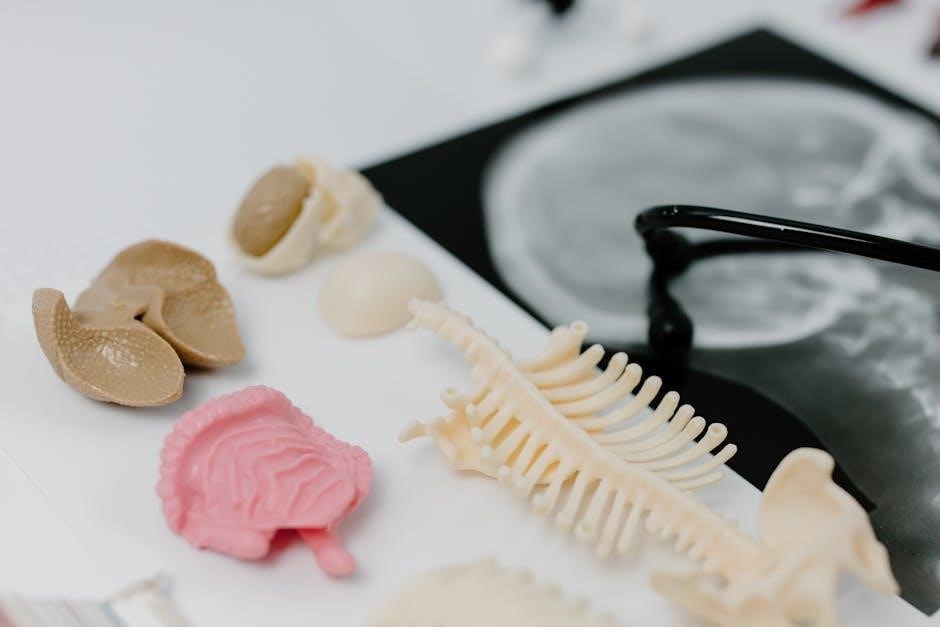
16.2 Diagnostic Techniques
Diagnostic techniques are essential for assessing physiological conditions and detecting abnormalities. Common methods include imaging technologies like X-rays, MRIs, and CT scans, which provide detailed internal views. Laboratory tests, such as blood and urinalysis, help identify biomarkers and infections. These tools enable accurate diagnoses, guiding treatment plans and monitoring health progress, bridging lab learning with real-world clinical applications.